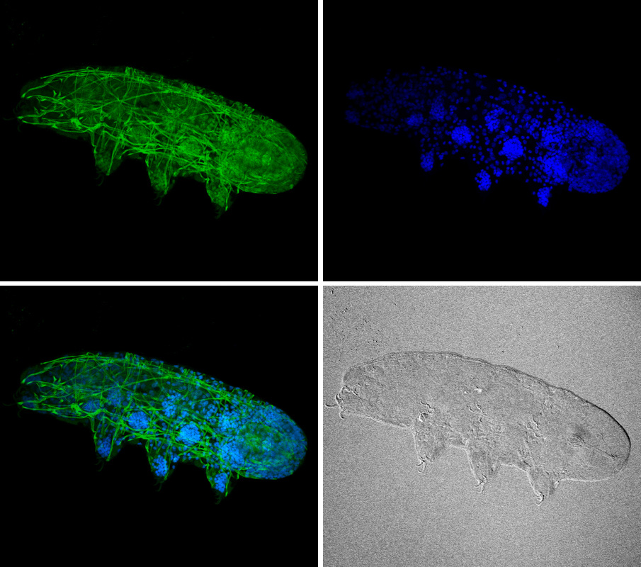
This week we have a submission from Dr. Frank Smith. Dr. Smith used confocal fluorescence microscopy to image this H. dujardini specimen. He used phalloidin (green) to label F-actin (muscles) and DAPI (blue) to label DNA. Frank used this and other techniques to study the segmental body pattern and brain architecture of this tardigrade species. You can read about his findings here. Be sure to check out Frank's movies lower in this post.
Below are two movies from Dr. Smith. The first shows a 3D model of the musculature of the tardigrade H. dujardini. The second shows a z-stack zoom through of a tardigrade with labeled muscles (green) and DNA (blue).
| | |
 RSS Feed
